“Beyond the X-Ray”
From David Rabkin 6/20/05
“The first phase of our exhibit on medical imaging – now titled “Beyond the X-Ray” – was opened to the public on early May! It’s a terrific start with some beautiful components. Many thanks to you for the help and advice offered by so many of you.”
The Common Vein contributed in two ways to the exhibit. Some of the images from the “Art in Biology” collection were displayed and those are presented below. They included “‘Chest of Fruit”, “Scaffolding of the Heart”, “Listen with your Eyes”, “Grapes of the Lungs”, “Emphysema”, and poetry and art surrounding the “Hermit of the Abdomen” – aka the pancreas
The second component was the involvement of the “Radiologist for the Day” – an interactive program emphasizing the evaluation of lung nodules. Patients of differing backgrounds were found to have lung nodules and the audience was asked to determine whether the nodules were of a concerning nature or whether they were benign findings.
The following images represent the exhibit of “Art in Biology”
Chest of Fruit
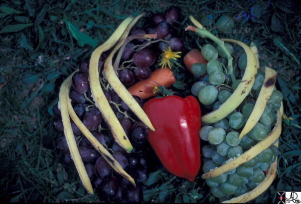
Chest of Fruit |
| This artistic rendition of the heart and lungs uses the shape of fruit and vegetables to create an image of the chest. The lungs are made of grapes, the pulmonary arteries are made of carrots, the ribs are made of banana peel and the heat is made of a red pepper. 02032p Courtesy Ashley Davidoff MD. accessory cardiac heart lung bone PA grape banana peel ribs Davidoff art |
Scaffolding of the Heart
The heart is built around a scaffolding that consists of a horizontal line that is bisected by a longer vertical line. In the example below the arms of my daughter are positioned in the horizontal projection along the expected position of the atrioventricular groove, while her legs are positioned along the vertical axis of the interventricular septum and her head is sitting in the vertical axis of the interatrial septum.
Scaffolding of the Heart |
| The scaffolding of the heart can be represented by a cross, consisting of two upper smaller components, and two larger lower components. This is only a conceptual 2 dimensional and symmetrical model that will help you understand the structure of the chambers and how the vessels and nerves are arranged around the scaffolding. As the story unfolds the complexity of structure will unfold, but it is important to understand that underlying the complexity there is a simple infrastructure. Courtesy Ashley Davidoff MD 32058 code cardiac heart introduction infrastructure drawing Davidoff art |
Listen to the Chest with Your Eyes
The key to excellent patient care is the combination of the clinical (symtoms signs) presentation and the application of the technology. The exchange of these two disciplines by face to face and personal communication of radiologist and clinician in this instance, provides the patient with the best care.

Listen with Your Eyes |
| When technology meets the art of clinical acumen, there is no more powerful tool in medicine. Communication between the imaging specialist and the clinician is key to excellent care. 32647 Courtesy Ashley Davidoff MD. accessory interesting Imaging Strategies Chest Stethoscope heart cardiac chest X-ray lung Davidoff art |
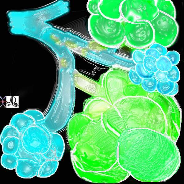
Grapes of the Lungs
The smallest pulmonary unit is the alveolus and the image below represents the cluster of grapes on the terminal and repiratory bronchioles
Normal Alveoli or Grapes of the lung |
| 32645a10.800 This diagram illustrates the branching pattern of the tracheobronchial tree that extends from the bronchi to the terminal bronchioles transitioning into the alveoli via the alveolar sacs. Courtesy Ashley Davidoff MD 32645b04b04 lung Davidoff tree branching alveolus alveoli normal drawing Davidoff art |
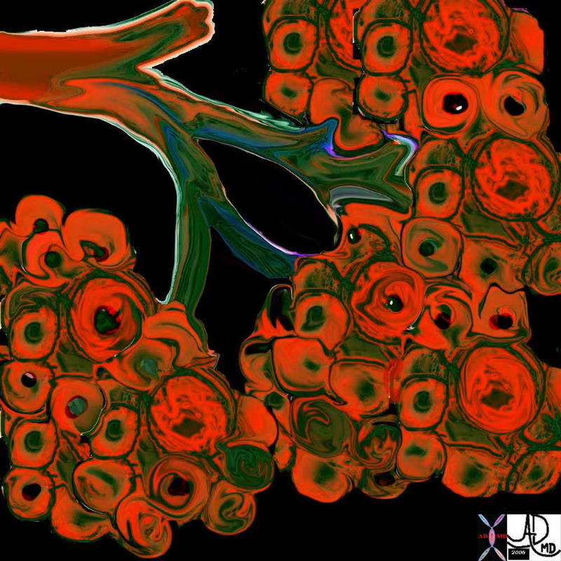
Emphysema
Loss of elasticity caused by destruction by elastic tissue causes the terminal airways the dilate hence trapping air. The image below captures the effect of smoking on the airways.
Emphysema |
| A drawing showing the normal acinus in teal and the abnormal emphysematous acinus in green characterised by destruction of the septal walls, emlargment of the alveoli, and loss of elasticity. The absence of involvement of the respiratory bronchiole makes the pathological diagnosis of centrilobular emphysema. Courtesy Ashley DAvidoff MD. 32645 |
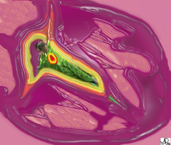
The Hermit of the Abdomen – a poem outlining the history of the pancreas.
Copyright 2007
Knowledge of the the pancreas eluded scientists for years. Modern technology has exposed this shy organ – The events of the history and evolution of the science and biology of this odd shaped organ captured my interest and inspired me to write a poem about it. The full extent of the poem was not published in the museum exhibition but I have included it here.
You have been called the hermit of the abdomen
By whom I do not know
But in your dark and hidden way, you have
spoken without a word
from the gurgling depths of the abdomen
Yes – you have earned this lonely title
and a coin should be tossed
to the person who coined the phrase
But it took a long time to understand who this hermit was – and what he was doing in the darkness of the abdomen
From the day of antiquity
You have been looked upon by many
Herophilus, the father of anatomy had the first incisive insights into you
As he was one of very few who had the guts to explore the guts in open fashion
Aristotle at the same time seemed to have known something about you
but then you lay unharmed and unexplored for almost 500 years
Until Rufus mistook you for a piece of meat –
You must have laughed at the “pan kreas” thing
How wrong he was – you evasive little trickster
and then the Talmud – always seeming to be right
thought you were the finger of the liver –
little did they know how independant you were
While Vesalius was up to your “hide and seek” game
the magical eyes of da Vinci missed you completely
even though he saw the serpiginous splenic artery snake right above you
Your ducts seemed to have intrigued the next generation Wharton Wirsung and de Graaf
As you sustained the pain of the quill penetrating your inner gut
(I forget you were already dead but it must of hurt just watching!)
A little later it was that man called Vater and the little Italian Santorini who found your minor duct and your nipple
And so by this time we had a good understanding of you in your nakedness
but of course, as said – you were dead
And so young Benard explored your factories, and got a sense of your canine workings,
But you were able to hold on to your sweet secret for just a little longer
until the Langerhans found the family jewels in the famous 2% of your population-
The islets – those beautiful eyelits – governess of all things sweet in the body
Eberle Bernard Danilevsky, and Kuhne joined up across the world to expose your antacid and enigmatic enzymatic brew
And once again your wonderful workings for a better world were exposed –
and we knew then, that you were the quiet and effective type –
a hermit who did good
but did not want the limelight
To see you as you lived and breathed in the flesh
was the mission of Wilhelm the X-Ray man
who crusaded the path to visualise 40,000 Angstroms under the skin
And then there was a slew of heroes who learned to slew your sickened parts – including the famous Whipple who was able to Whipple you in an inimitable way
And then a bone guy – for God’s sakes – a bone guy! – called Banting and his student Best
exposed the insular chemistry of you insulin that had given you the power over the sweet
Never mind – in the end it was for the good of all –
And a new era was borne
And so we try to understand your form as our scans explore you as you live and breathe
and we stare in awe at your odd shape – why oh why did you choose that shape?
What are you supposed to look like? – we have no clue
And we are happy – so happy for you that you are well nourished by a double blood supply
And we wonder why you have no skin – we thought all the organs had a skin
except for your tail – almost a foreskin
And you are off axis on two planes – what is that all about? – kinda crooked
And your twin origins and the intimacy with the duodenum, of the ventral twin
And the strange fusion of the Wirsung guy excluding the little Italian Santorini
It seems to me that your matrimonial fusion with Wirsung and the bile duct has led to more problems than the merger was worth
It does not seem in the long run, to have been a marriage made in heaven
What was that all about? Is there a grand plan to come
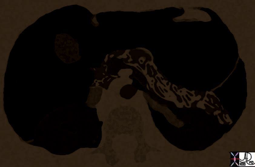
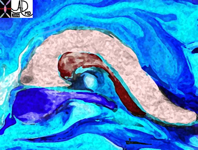
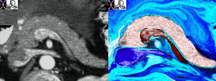
Pancreas – Coming Out Party |
| 38025b10 Courtesy Ashley Davidoff MD code pancreas pancreatic shape code liver code stomach code kidney renal code lesser sac code tuber omentale code lesser omentum drawing anatomy normal Davidoff art |
And so we try to understand your diseases
And in some way we understand that the guy glugging down the bottle
Could be punished by your reaction
But why Oh why are you so nasty to those whose misfortune it is to have stones roll down and get no satisfaction.. down the green vile bile route
Have you not learned to live with the green secretion by now
And did you not know that by reacting the way you do, that you are cutting off your nose to spite you head?
While type 2 seems remote from you
we don’t know about this Type 1 business
why are you made to suffer so much at the hands of your own body on your own body
we feel sorry for you – to have your own buddys reject you – must be awful
and then to see so many young ones suffer because you dont work
and we once again see and understand what power you control from that deep dark hermit home of yours
And the cancer thing … so silently it creeps on you causing your collagen to counter
and only making things worse as it strangles nerve, blood vessel, and your spouse duct – the green one, – without regard
And then I think of you in your prime and in your happiness
When you are with you two buddies – the splenic vein and the renal vein
And you all look so much alike, and happy swimming in that deep ocean where you hide
And I wish this was forever
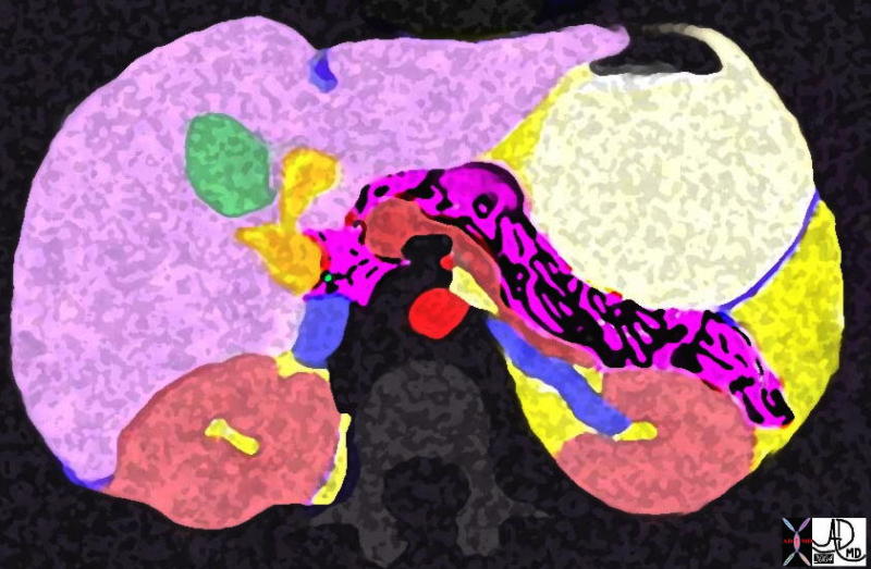
Pancreas with his Buddies the Splenic Vein and the Left Renal Vein |
| 24796b07 Courtesy Ashley Davidoff MD pancreas pancreatic anatomy normal relations splenic vein SMV IVC renal vein kidney relations Davidoff art |
Planning the exhibit “Beyond the X-Ray”
extracts from an email from David Rabkin 6/21/04
“The two main categories will be “intriguing” images and good examples of diagnostically useful images.”
“The exhibit will use images of everyday objects selected by participating radiologists (flowers, fruit, appliances, dolls) to draw in visitors and arouse curiosity. We know that some of these images are gorgeous and so here may be a bit of an art gallery feel to this portion of the exhibit. By adding radiologists’ interpretations of the images, museum-goers will get a sense of what the different imaging technologies actually do. That creates the opening for us to tackle how they work, what the patient’s experience is like, considerations doctors use in selecting which technologies to use and when, and how they’re actually used diagnostically. At the Museum we use a lot of live interpretation; so we aim to have a series of guests from the radiology community as well as our own staff interpreting on the floor.”
Reply fromDavidoff 6/23/04
“Thanks for the invitation – I have a large collection of images – both medical and creative/artistic – If it is creative and involves images -count me in.”
Email from Davidoff 030505
“There are a few aspects that I am personally excited and inspired about – the detective stuff – as discussed – there are some great Sherlock Holmes quotes that go with the flow “not invisible but unnoticed – you did not know where to look so you missed all that was important” or something like that
…and the artsy stuff – I have created both poetry and art around imaging the body Recently (and it is still in progress) the pancreas has grabbed my fancy – the pancreas eluded us for so many years hiding in a deep dark pocket of the belly CT has exposed it it in all it’s glory – it used to be called the hermit of the abdomen – no longer
There is a marvelous way that one can tie the art of looking at a radiologic image and looking at, and seeing a painting – in essence the art of looking and the science of seeing (“a picture speaks a thousand words – but you need to know the thousand words” Please excuse the rapid fire of ideas – I think, the creation of your exhibition and a forum and platform for our profession is the stimulus
From David Rabkin 04 04 05
“I know the team is really excited about your involvement and really appreciated the visit last week. I’ve got to find out what they are talking about when they referenced your “pancreas poetry and art” and I very much hope we develop an ongoing working relationship that you’re completely comfortable with.”
from David Rabkin 4/25/05
“We really need you to be an advisor, not a designer. I know it’s tough given your artistic inclinations and your plans for doing education work in this realm. I developed many of the early ideas that defined the project and at times find it really difficult to let go when the team heads in a different design direction or feels they can’t fit in something that interests me. But I’ve learned to trust this group and their expertise in museum-based education; we all must.”
From David Rabkin 6/20/05
“The first phase of our exhibit on medical imaging – now titled “Beyond the X-Ray” – was opened to the public on early May! It’s a terrific start with some beautiful components. Many thanks to you for the help and advice offered by so many of you.”
Radiologist for The Day
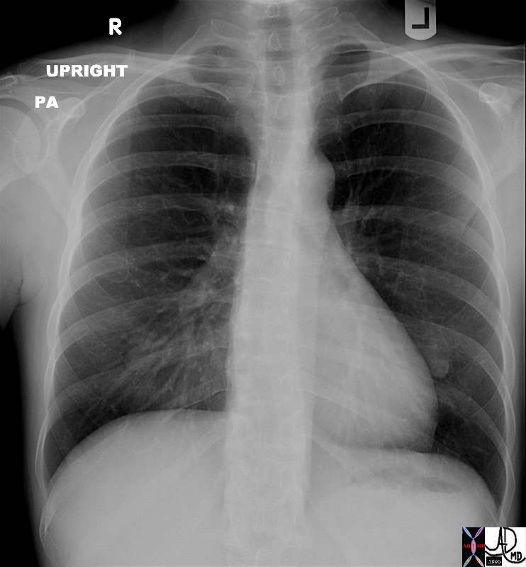
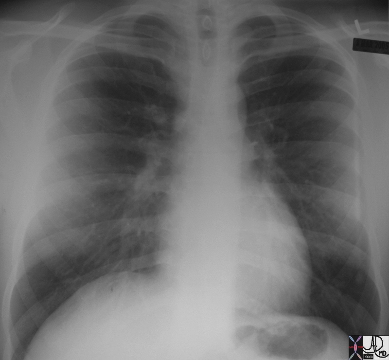
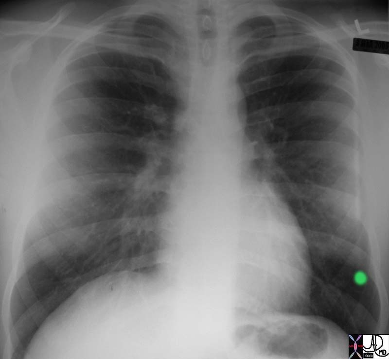
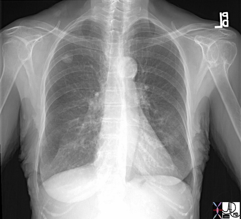
Normal Chest |
| 41819c Courtesy Ashley Davidoff MD medical students code chest imaging radiology plain film CXR lung normal |
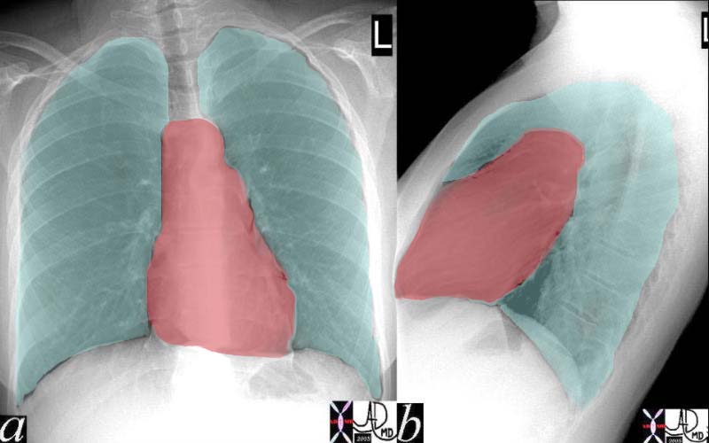
The Heart and the Lungs |
| 41819c04 normal |
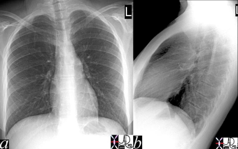
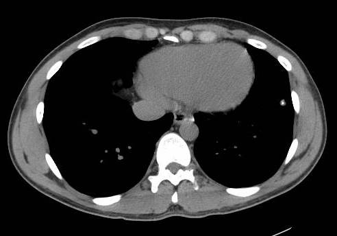
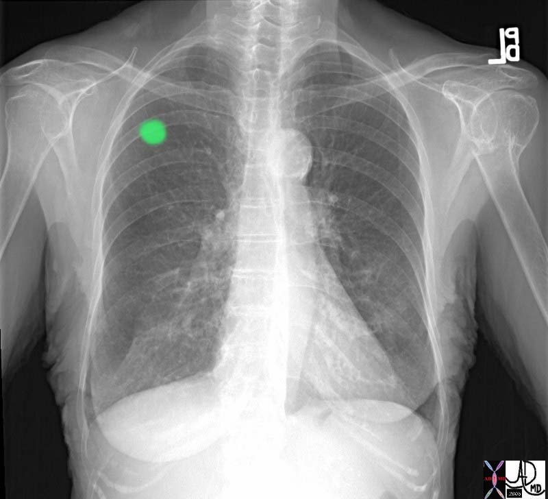
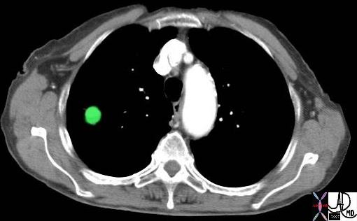
Lung Nodules
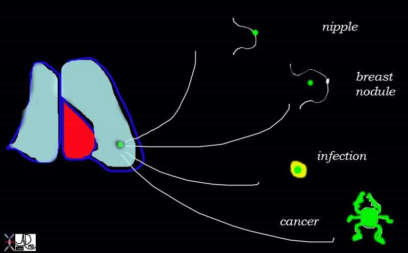
Some Types of Lung Nodules |
| 42274b01 Courtesy Ashley Davidoff MD RS lung fx lung nodule MOS medical students |
 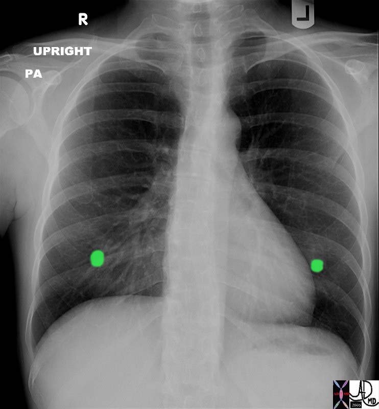
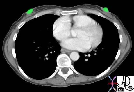
Bilateral Nodules – Breast Nipples |
| 42252 Courtesy Ashley Davidoff MD RS lung fx lung nodule MOS medical students |
42254 |
| 42254 Courtesy Ashley Davidoff MD RS lung fx lung nodule MOS medical students |
 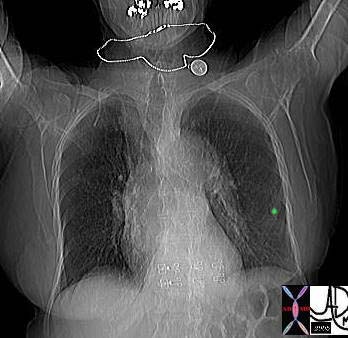
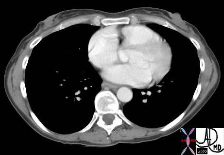
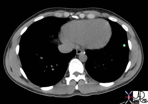
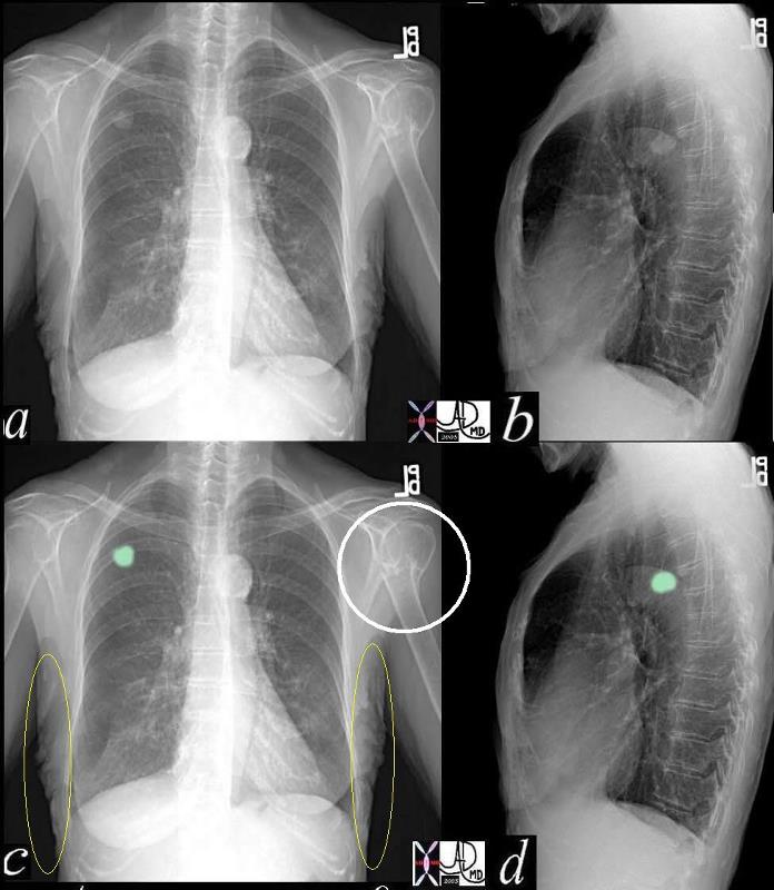
Left Lower Lung Nodule |
| 42245 Courtesy Ashley Davidoff MD RS lung fx lung nodule MOS medical students |
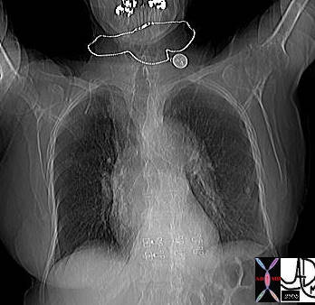
Calcified Breast Nodule |
| 42247 Courtesy Ashley Davidoff MD RS lung fx lung nodule MOS medical students |
  Left Lower Lobe Nodule Left Lower Lobe Nodule |
| 42235 Courtesy Ashley Davidoff MD RS lung fx lung nodule MOS medical students |
Central Calcification – Benign |
| 42243b01 Courtesy Ashley Davidoff MD RS lung fx lung nodule MOS medical students |
Central Calcification – Benign |
| 42244c01 Courtesy Ashley Davidoff MD RS lung fx lung nodule MOS medical students |
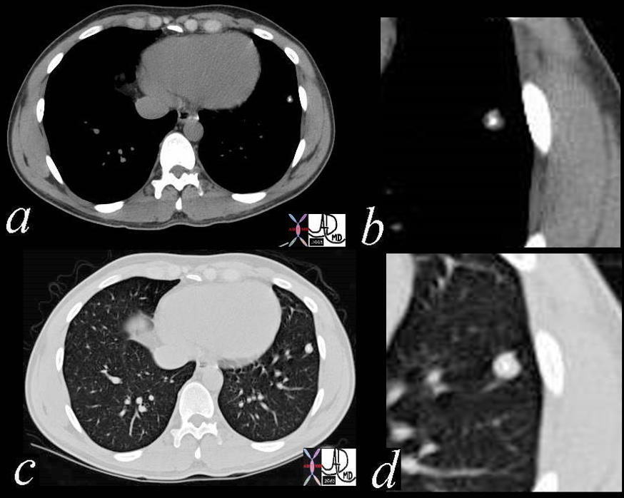
  The Lung Nodule The Lung Nodule |
| 42262 Courtesy Ashley Davidoff MD RS lung fx lung nodule MOS medical students |
Other Findings |
| 42263c03 Courtesy Ashley Davidoff MD RS lung fx lung nodule MOS medical students |
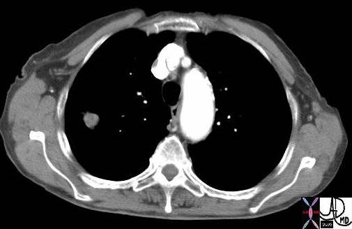 
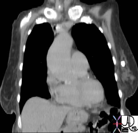
Lung Nodule – CTscan |
| 42269 Courtesy Ashley Davidoff MD RS lung fx lung nodule MOS medical students |
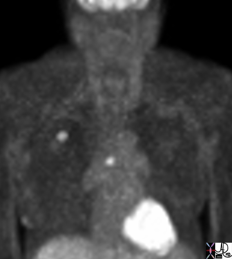 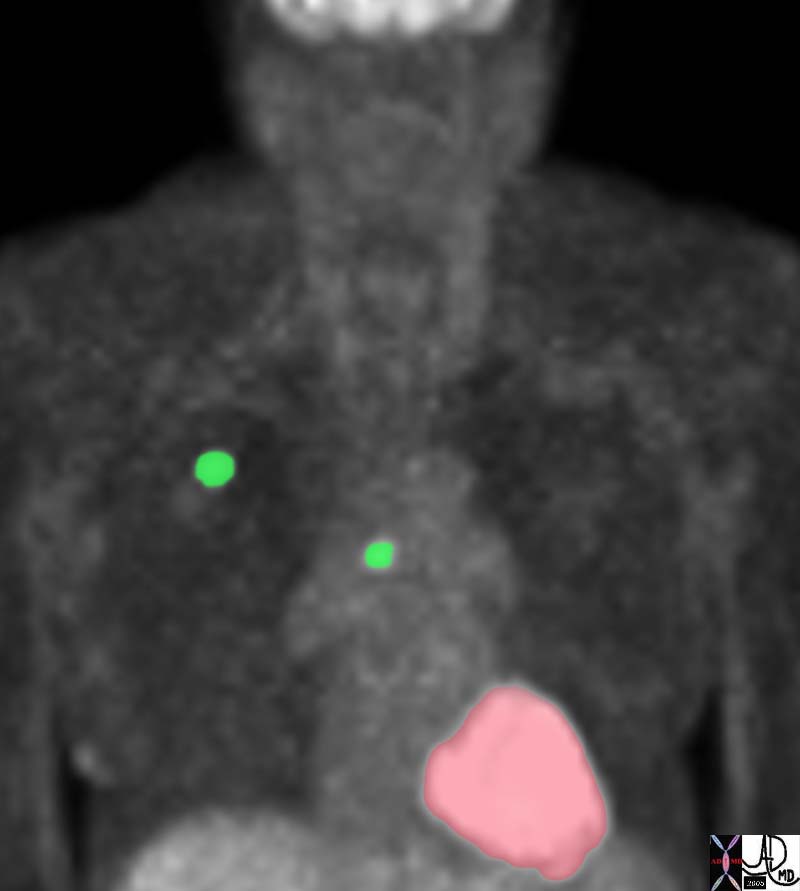
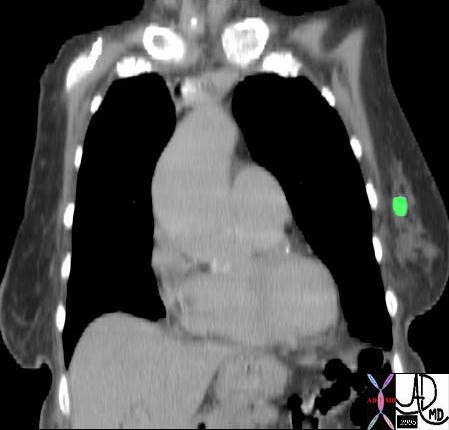
Concerning Lung Nodule – Positive PET scan |
| 42271b Courtesy Ashley Davidoff MD RS lung fx lung nodule MOS medical students |